 1.
Step/Stride length
1.
Step/Stride length
(Click on the images to see the movies. The introductory notes are here)
1.
Step/Stride length
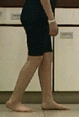
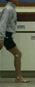
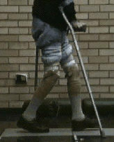
The most important functional measure. Calculate it using the formula:
Stride length = (120 x Velocity)/cadence.
Remember that One Stride = 2 Steps (so step length = stride length / 2).
Normal is 1.5 m for an adult, and approximately equal to 0.9 x height (stature) for a child.
This child's step length is obviously too short, compared to her height.
Usually causes: inadequate push-off/pull-off, pain.
 2.
Ankle angle at contact
2.
Ankle angle at contact 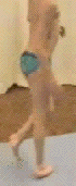
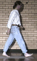
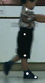
The ankle should be close to it's neutral position (a right angle) when it strikes the floor (left picture/video).
It can be plantarflexed for two reasons:
1. Equinus deformity (Triceps, especially gastrocnemius spasticity), or
2. Drop foot (flaccid paralysis of pre-tibials, which normally lift the foot in terminal swing)
Both cause the patient to make a forefoot contact instead of the normal "heel-strike. The child on the right has an equinus deformity from cerebral palsy diplegia.
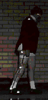
 3. Ankle angle at toe-off
The push-off produced by the triceps surae (gastrocnemius + soleus) is the main muscle group responsible for causing the leg to swing forward (swing leg advancement).
When there is weak push-off (often called an apropulsive gait), the stride length will be reduced, and gait velocity falls.
This child has had a common operation for equinus deformity, in which the Tendo-Achilles is lengthened.
This fixes the equinus deformity, but unfortunately very often reduces push-off power becuase the muscles are now too long to work effectively.You can see that she has lost the normal plantarflexion during pre-swing.
 4. Knee angle at contact
The knee should normally be close to full extension at initial contact.
This is important to achieve a correct loading response, when the knee is allowed to flex slightly (stance phase flexion) under eccentric quadriceps control.
In this child, the knee is at an angle of nearly 30 degrees at contact, and in the video you can see that this makes his loading response very abnormal.
Note also that he lands flat-footed - this is a natural consequence of the excessive knee flexion.
Common causes are a knee flexion contracture, or hamstrings tightness due to a muscle sprain.
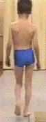
 5.
Amount of stance phase knee flexion
5.
Amount of stance phase knee flexion
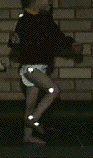
The small amount (about 20 degrees) of flexion at the knee during stance phase is one of the determinants of gait. It helps to improve energy efficiency by lowing the body centre of gravity during stance phase, when it is at it's highest.
This child (who has Down's syndrome) has lost the normal stance flexion, and has a fully extended knee throughout stance.
Probable cause: quadriceps weakness. When the quadriceps is not strong enough to eccentrically control the knee during loading, the plantarflexor-knee extensor couple is used instead.This requires body weight to be brought forward in front of the knee joint axis. Patients with knee arthritis also use this mechanism to reduce pain because passive stabilisation produces lower bone-on-bone forces than use of the quadriceps.
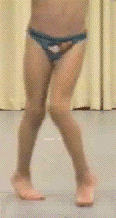
 6.
Amount of swing phase flexion
6.
Amount of swing phase flexion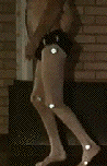
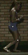
Normal swing-phase knee flexion should be around 65 degrees at normal cadence. At lower speeds it is much less than this (try walking at a very low speed, and you will find you walk with an almost straight leg).
This baby's swing knee flexion is excessive (around 90 degrees).The knee is hyper-mobile.
In contrast, the man on the right has a fixed knee deformity from spastic contracture. His knee does not flex normally in swing - in fact it stays fixed at the same angle (about 30 degrees) all the time.
 7.
Hip extension in terminal stance
7.
Hip extension in terminal stance
The hip should normally extend about 20 degrees in terminal stance.
If the hip fails to extend, a common cause is a flexion contracture of the hip - a frequent complication of sitting a wheelchair for too long.
This man had peripheral vascular disease from smoking, and has had to have both his legs amputated.
He's now learning to walk again with bilateral below-knee prostheses, and has obvious hip flexion contractures.
This should be confirmed by examining the patient in supine lying, with a hand behind the lumbar spine to check for lordosis (Thomas test).
 8. Trunk angle (forward flexed?)
A forward lean of the trunk is a common compensation for a short stride length, so it can be seen in many pathological gaits.
Flexing the trunk helps in two ways:
1. By placing body weight ahead of the knee joint, it stabilises it with the knee extensor-plantarflexion couple mechanism.
2. It aids flexion of the hip (pull-off), by moving the body weight (ground reaction) line anterior to the hip.
This patient is using a long leg brace (splint) on her leg, which is weak from poliomyelitis. She leans her trunk forwards in pre-swing because her hip flexors are weak and she has no ankle push-off.
 9. Frontal plane: Trendelenberg Sign
This is best looked for from behind the patient.
The hip abductors normally support the pelvis during swing. When they are weak, the pelvis drops on the swing side.
The overall effect is of a "waddling" gait, like a duck!
One problem it causes for the patient is that the clearance is reduced, so they may compensate by circumduction or vaulting with the opposite ankle.
This sign is common in many pathologies, including hip osteoarthritis, stroke and cerebral palsy.
![]() 10. Transverse plane: angle of patellae & feet, and arm posture
10. Transverse plane: angle of patellae & feet, and arm posture
Looking from the front, notice that the patellae of this child, which should normally face forwards, are rotated inward. Note also that the feet are turned inwards (adducted). These problems indicate transverse-plane (rotational) abnormalities (called "femoral anteversion and/or tibial torsion").
The internally rotated feet will tend to reduce efficiency, because the moment arm of the triceps muscle during push-off will be reduced - so-called lever-arm dysfunction.
Don't forget the arms too - the posture of the left arm of the child on the right gives a clue that something is wrong on that side. A close look reveals the patella to be rotated slightly externally.

Dr. Chris Kirtley, Associate Professor, Dept. Biomedical Engineering, The Catholic University of America, Washington DC